Visited the dentist, had your teeth treated and after a while it started bothering you again? The cause may be secondary caries under the filling of the tooth. The situation is unpleasant, but quite common. Unfortunately, for a long time the carious process is asymptomatic. Patients begin to notice that the filling has changed color, broke off, the tooth began to react to temperature stimuli, when the disease has developed to a deep stage.
Causes of secondary caries under the filling
The main cause is the same as in primary caries: the penetration of bacteria through the protective barrier. This barrier in the treated tooth is the filling. As soon as its integrity is compromised, problems arise. Bacteria are able to penetrate a microcavity 50 microns wide. They begin to multiply, eating away at the hard tissue.
Factors provoking secondary tooth decay:
- improper bite;
- poor oral hygiene;
- weakened immune system;
- bruxism;
- poor quality of the filling.
Composite fillings tend to shrink. In addition, fillings can not withstand overload, so dentists urge not to chew nuts, candy and seeds. Filling materials do not like sharp temperature changes, for example, when a hot cake is drunk with an ice-cold drink.
Deep caries under the filling is also formed as a result of violation of filling technology:
- Not all the diseased tissue was removed. Part of it, and therefore bacteria, remained under the filling and continue their destructive work.
- Did not use liquid-fluid polymers for better adhesion (gluing, soldering) of materials.
- The photopolymer filling was not applied layer by layer, but in one mass. Such a filling quickly gives shrinkage, opening the way for pathogens.
- The filling was not polished. Roughness and bumps serve as an excellent landing pad for microflora. Plaque gradually forms, and then caries.
Another reason is that fillings do not last forever. Old fillings wear out, even if you follow all the requirements of technology. The service life of the filling material, and most importantly, the adhesive, 5-7 years. After that, it is advisable to change the filling.
What is the danger of secondary caries under the filling?
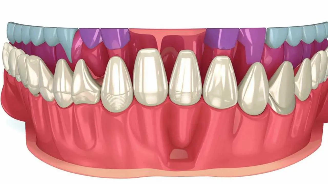
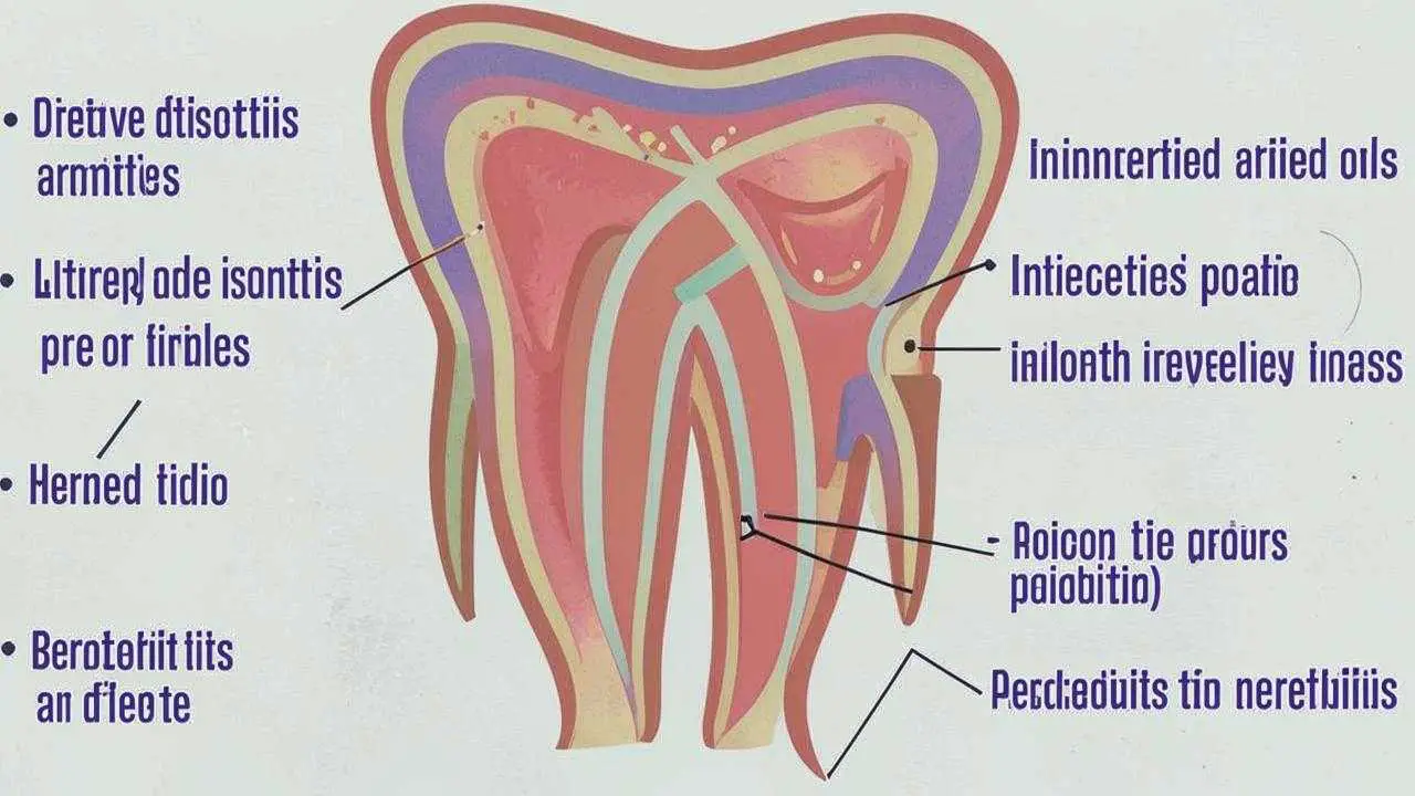
Caries is always associated with demineralization of enamel. This means that the teeth begin to react to any stimuli, hypersensitivity develops. The protective properties of enamel are reduced, an increasing number of pathogens penetrate into the deep tissues. They provoke pulpitis – inflammation of the pulp (nerve) or periodontitis – inflammation of the root.
Pulpitis
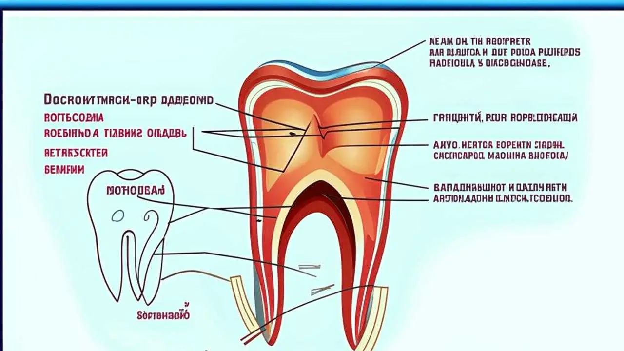
At first, the disease affects not the entire nerve-vascular bundle, but only part of it. During this period, the tooth can be treated without removing the pulp. But the development of the disease occurs very quickly and in a few days affects the entire pulp. The patient begins to experience severe pain. In such cases, the nerve has to be removed, the depulped tooth becomes more fragile. If pulpitis is not treated in time, pus begins to accumulate in the pulp chamber, from it bacteria get into the general bloodstream, causing inflammation in the joints and bones of the jaw. Opening the mouth becomes a problem, let alone chewing. If the inflammation has spread deep into the tissues, the tooth will have to be extracted.
Periodontitis
Inflammation of the tooth root and surrounding tissues is a complex pathology. Treatment is endodontic, with washing and filling the canals. The constant inflammatory process, purulent masses in the tissues lead to the fact that the infection affects the internal organs, in particular the heart. The connection between periodontitis and heart disease is confirmed by scientific research [1].
It is in periodontitis that flux develops – a pronounced swelling of the gum caused by pus accumulation. In some cases, a cyst forms at the root, which is surgically removed.
Other complications of secondary deep caries can be phlegmons (inflammation of soft tissues) and osteomyelitis (inflammation of the bones of the jaw). These are dangerous to general health and painful pathologies.
Diagnosis and treatment
To determine that caries has developed on a filled tooth, dentistry uses the following methods:
Patient complaints that the tongue feels chipped, gaps, the tooth hurts, hypersensitivity has developed, help the doctor to make the correct diagnosis.
Changes in the color of enamel, the appearance of swelling, swelling indicate possible infection.
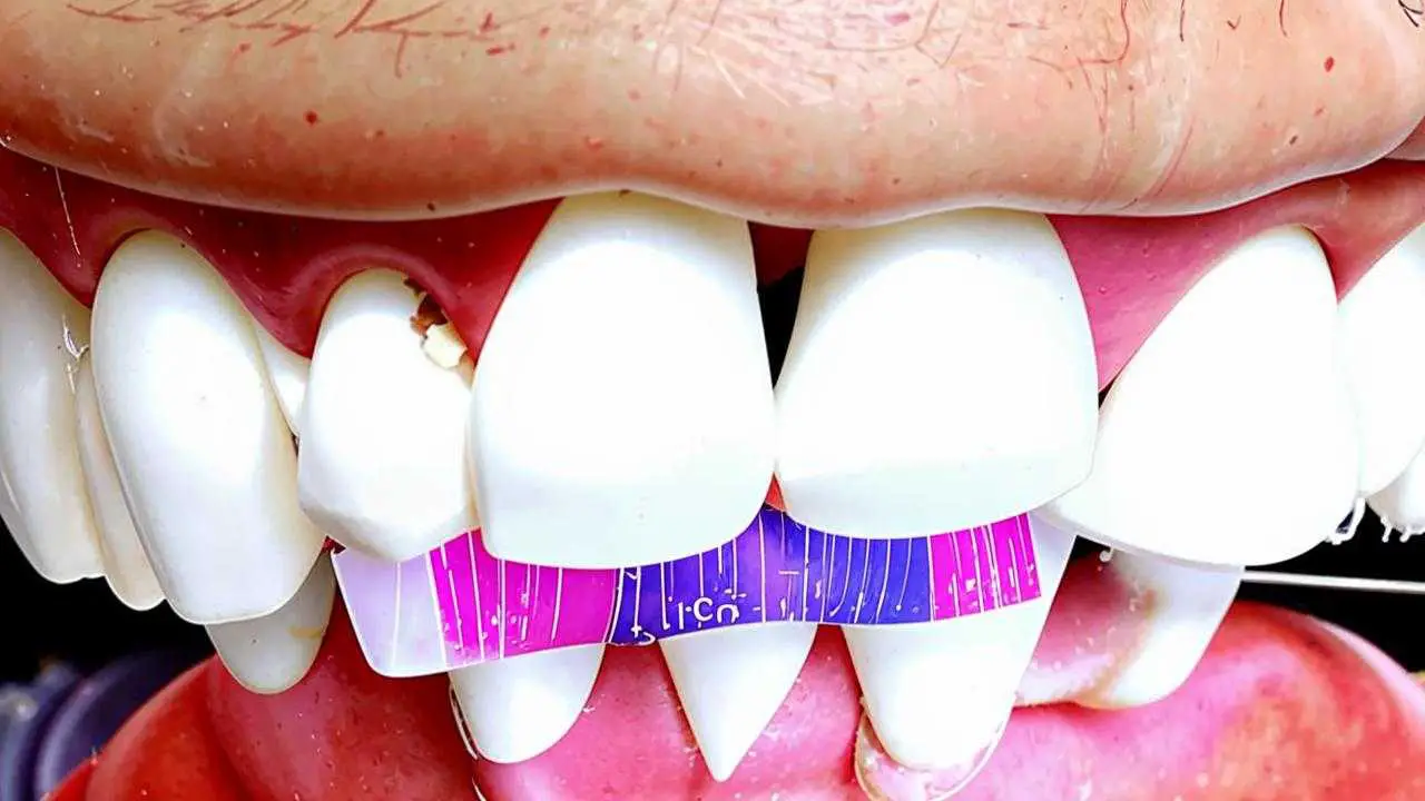
The probe gets stuck in the gaps, which already indicates the possibility of carious lesions due to the violation of the integrity of the filling.
- Vital staining
Do if the signs of caries are not obvious. A special solution stains the tissues affected by infection. This method allows you to diagnose caries, even if there are no symptoms.
The doctor records the reaction of the tooth to warm and cold air or water.
- X-ray examination
Determines the caries under the filling on X-ray, the degree of lesions, the presence of granulomas, periodontitis.
- Electroodontometry
An electrical signal causes the pulp to react. Helps diagnose pulpitis.
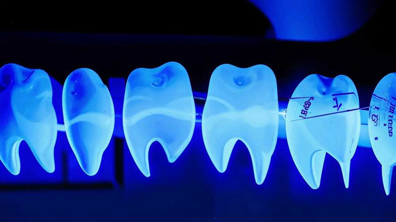
- Luminescence
When exposed to UV light, infected tissue changes color. This is the main sign of tooth decay.
Treatment of secondary caries
In each clinical case, a different treatment plan is drawn up, it depends on the location of caries, the depth of penetration, the area of spread.
If the focus is localized, the pulp or root is not affected, it is enough to replace the filling, removing the affected tissue.
If the inflammation has spread to the pulp, it is removed, treated and filled canals, and then seal the carious cavity.
In periodontitis, surgical methods are used. The specific operation depends on the type of inflammation. It can be resection of the root tip, hemisection (removal of one outgrowth), separation or removal of the cyst. Only after that, canal filling is performed and the filling is restored.
After endodontic (canal treatment) intervention, prosthetics with crowns or inlays are recommended.
Профілактика
To avoid tooth decay on a filled tooth, the dentist should make sure that:
- all diseased tissue from the treatment of primary caries is removed;
- the filling fits snugly against the tooth walls;
- the surface of the filling is polished, smooth, without gaps.
The use of modern, high-quality materials such as glass ionomers significantly reduces the risk of re-infection.
In turn, patients should also take measures to prevent a situation where a tooth has rotted under a filling. To do this, it is important to:
- Brush your teeth at least twice a day;
- floss;
- Have regular professional dental cleanings.
If you notice that the tooth enamel has changed color, cracks and chips have appeared, the gum is swollen or the tooth reacts to temperature stimuli – do not delay a visit to the clinic. A timely diagnosis and treatment can help you avoid complications.
Sources:
[1] https://minzdrav.gov-murman.ru/documents/poryadki-okazaniya-meditsinskoy-pomoshchi/6_periapikal.pdf
