Necrosis of the jaw is a severe inflammatory disease in which the bones of the facial skeleton are exposed and die off.
Cellular die-off is provoked by:
- Radiation therapy. Moreover, necrosis of the jaw bone can develop some time after the end of radiation, which makes diagnosis difficult.
- Intake of synthetic drugs. Drugs destroy osteoblasts – the basis of bone tissue.
- Taking bisphosphonate drugs. These drugs are prescribed for osteoporosis (in small doses) and oncologic diseases that affect the bones of the skeleton (long-term, in large doses).
- Decreased immunity as a result of diseases (rheumatism, blood diseases, polyarthritis, diabetes) or chemotherapy.
- Infectious diseases – general and oral.
- Injuries to the jaw.
Causes of jaw bone destruction
The disease develops when several factors combine. With other identical conditions, patients fall into the risk group:
- with poor oral hygiene;
- with periodontitis;
- after dental surgery, including tooth extraction;
- taking large doses of bisphosphonates for a long time;
- during chemotherapy and corticosteroid therapy;
- undergoing treatment with antineoplastic drugs;
- with diabetes;
- with ill-fitting, chafing dentures.
Infections of any etiology greatly increase the possibility of becoming ill. Untreated, carious teeth open the way to pathogens not only in the oral cavity. In neglected cases, when purulent discharge begins, the infection enters the general bloodstream and spreads throughout the body. On the other hand, sinusitis, otitis, sore throats are a huge number of pathogens that provoke serious dental diseases. Complication in both cases can lead to osteomyelitis, and he – to necrosis of the jaw.
Symptoms and course
Unfortunately, for a long time the disease can proceed without symptoms at all. The patient does not feel pain, so the doctor turns to the doctor when the infection has penetrated deeply into the bone.
Signs of necrosis of the jaw:
- pain in the tooth affected by the infection or in the hole of the extracted tooth;
- loosening of the teeth;
- swelling that is so severe that it disrupts facial symmetry;
- purulent discharge from the gums;
- putrid breath odor;
- painful sensations when swallowing, chewing, talking.
The main sign of osteonecrosis of the jaw is the exposure of the bone.
Classification
The course of the disease, provoked by infection can have an acute, chronic and subacute character.
According to the location, there are distinguished:
- osteonecrosis of the lower jaw;
- necrosis of the upper jaw.
By type of infection:
- endogenous – infection occurs as a result of dental diseases;
- Hematogenous – pathogens get into the jaw with the bloodstream.
Distinguish also:
- medicinal (bisphosphonate);
- traumatic.
The American Dental Association defines the following stages of the development of the disease:
Stage 0 – toothache that radiates to the temporomandibular joint or sinus. Tooth mobility, with preserved periodontium. Appearance of fistulas, cysts, fluxes.
Stage 1 – exposure of bone without severe pain.
Stage 2 – exposure of the bone area, accompanied by pain and inflammation.
Stage 3 – exposure of the bone area, which extends beyond the alveolar (the one in which the tooth is located) bone. With mandibular necrosis, the jaw is affected down to the lower edge. Bone atrophy occurs. This is the stage of pathologic fractures. With necrosis of the upper jaw, the zygomatic arch and maxillary sinus are affected. Numerous fistulas are formed.
Diagnosis
Symptoms of osteonecrosis of the jaw are similar to other diseases: tumors, bone tuberculosis, actinomycosis. Therefore, the examination is carried out comprehensively:
- Collection of anamnesis
If the patient has had osteomyelitis, takes large doses of bisphosphonates, underwent radiation therapy or had a trauma to the jaw, the doctor will conduct an additional examination. This will confirm or deny the presence of necrosis.
- Dental examination
If the bone is exposed, then the diagnosis is easier to make, but if the disease is at the zero stage, additional data are needed.
- Laboratory tests
Positive tests for c-reactive protein, leukocytosis, high COE – reasons to suspect a necrotic process. Also check the urine for the presence of protein, blood cells. In some cases, make an analysis of purulent discharge to determine the pathogen before prescribing bacterial therapy.
- Radiography
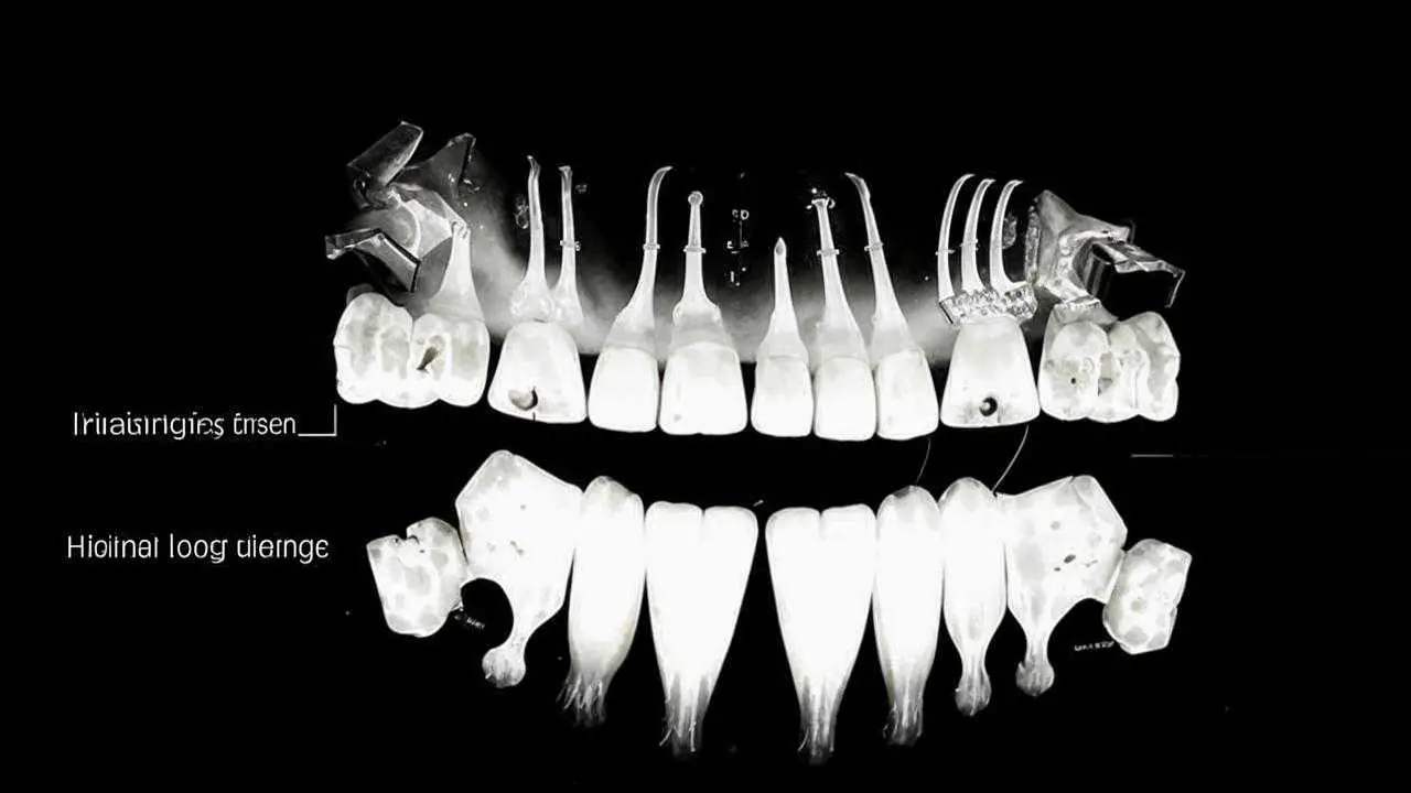
X-ray, or better CT (computed tomography) helps to determine the area of the pathologic process, the degree of lesion, the presence of fractures, sequesters (dead bone fragments lying freely between healthy tissues).
Treatment
The task of the dentist is to stop the destruction of bone tissue, prevent sepsis, relieve symptoms.
For this purpose, therapeutic and surgical methods are used, such as:
- Drug therapy
Prescribe broad-spectrum antibiotics, antihistamines, aseptic solutions for mouthwash. Analgesics are prescribed for pain.
- Surgical intervention
The doctor conducts curettage of pockets or wells of extracted teeth. He removes sequestrations, opens and provides drainage of purulent foci, splints mobile teeth.
Depending on the stage of the disease, surgery is indicated, in which the affected bone is removed completely, leaving only its healthy part.
In many cases, it is necessary to resort to reconstructive surgery to fill the defect or correct the deformity. This may involve bone grafting or jaw reconstruction.
The treatment of jaw necrosis is a multi-step process that requires the involvement of several doctors. Depending on the nature of the disease, the dentist works closely with an oncologist or general practitioner. For reconstructive surgeries under general anesthesia, the participation of an anesthesiologist and an oral surgeon is required.
Prevention
Timely dental care can prevent the development of purulent processes, the spread of infection to bone tissues. It is important to carry out sanitation of the oral cavity without delay, in order to destroy all foci of infection in time.
Doctors recommend eliminating all dental problems before starting treatment with bisphosphonates. If treatment is carried out during BF therapy – be sure to warn the doctor. He will take additional measures for antiseptic treatment, suture the wells, prescribe antibacterial therapy.
A healthy lifestyle, a balanced diet strengthens the immune system, does not allow infections to spread. This prevents the development of inflammatory processes and their complications in the form of necrosis of the jaw.

